-
- Global AMC MENU
- NEWS
- HEALTH
- PEOPLE
- Introduction

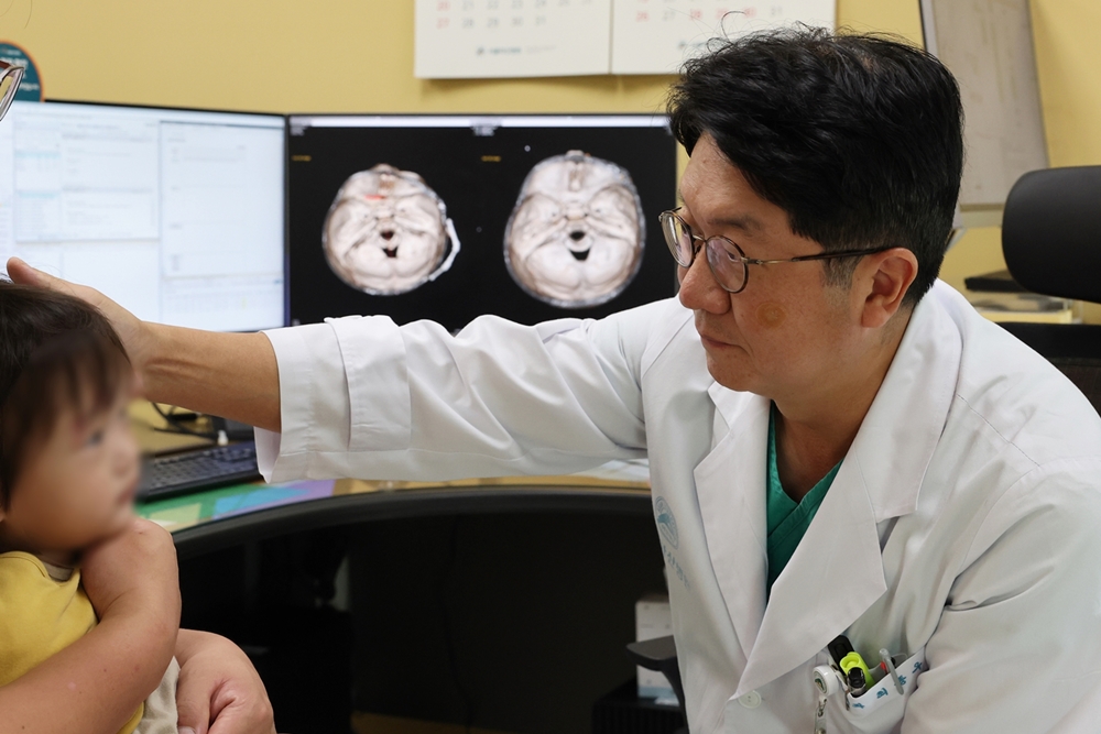
▲ Professor Jong-Woo Choi examines a pediatric patient who received cranioplasty using a distraction device.
Craniosynostosis is a rare condition where the skull bones join together early, blocking brain growth. Conventionally, it has been treated with surgery involving fragmenting and repositioning the skull bones. In 2005, the Craniofacial Clinic team, led by Professors Jong-Woo Choi and Young-Chul Kim of Plastic Surgery and Professors Young Shin Rah and Sangjoon Chong of Pediatric Neurosurgery, developed cranioplasty using a distraction device instead of the traditional surgery. Since then, the technique has safely treated about 140 children with craniosynostosis over 20 years, demonstrating its long-term effectiveness.
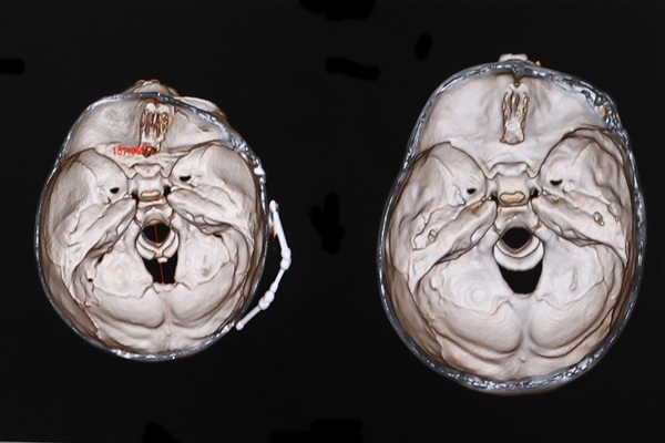
Cranioplasty using a distraction device does not involve fragmenting the child’s skull. Instead, a partial incision is made along the fusion of the cranial suture line, and the distraction device is attached. As the parents rotate the device 0.5 to 1.5 mm daily, the gap in the skull is expanded slightly for new bone to grow in the gap. Once the skull expansion has reached the normal range, the device is removed to complete the treatment.

▲ CT image of cranioplasty using a distraction device, gradually widening the bone area as shown on the left.
The technique has reduced surgery time by more than half, from about 8 hours to 3 hours, and the amount of blood loss has also significantly reduced. Scarring and complications are minimal, making it even safer, and the device-wearing period is shortened from 3 months to 2 months, allowing for faster recovery. Most of all, brain damage is nearly non-existent.

▲ (from the left) Professors Jong-Woo Choi and Young-Chul Kim of Plastic Surgery and Professors Young Shin Rah and Sangjoon Chong of Pediatric Neurosurgery
The average age of patients who underwent surgery was 10 months, with no deaths from the surgery. The skull grew symmetrically without recurrence in approximately 98% of patients, and the incidence of major complications, such as seizures or developmental delays, was 0%. Minor issues, including delayed wound healing and cerebrospinal fluid leakage, occurred in only 3% of cases and were successfully managed with conservative treatment. Both external and basal brain asymmetries were corrected immediately after surgery, with a long-term follow-up over 10 years confirming symmetrical facial bone development even after the patients grew older.
Over the past 20 years, distraction osteogenesis has appeared in more than 10 SCI journals, including the ‘Plastic and Reconstructive Surgery’ by the American Society of Plastic Surgeons. Recently, the technique has gained popularity in the United States and Europe.


