-
- Global AMC MENU
- NEWS
- HEALTH
- PEOPLE
- Introduction

▲ Professor Jun Ki Kim of the Department of Convergence Medicine at the University of Ulsan College of Medicine and Asan Medical Center
A research team led by Professor Jun Ki Kim of the Department of Convergence Medicine and Dr. Saeed Bohlooli Darian at Asan Medical Center has successfully captured real-time mitochondrial dynamics within the cells of small animal organs using an ultra-high-resolution imaging technology.
Mitochondria are extremely small organelles, nearly at the resolution limit of optical microscopy, and their subtle movements caused by respiration and heartbeat in living organisms make long-term imaging difficult. To overcome these limitations, the research team stabilized the imaging area by applying tissue fixation techniques to a two-photon intravital microscope. They enhanced image quality using a self-supervised learning-based noise-reduction model and improved resolution and clarity with the Super-Resolution Radial Fluctuations (SRRF) algorithm. Fourier Ring Correlation (FRC) analysis confirmed that the resolution of the processed images was improved by two to three times compared to the original.
When this technique was applied to a mouse model of alcoholic liver disease (ALD), real-time observation revealed dynamic changes in the mitochondrial network as the disease progressed. In addition, the administration of berberine, an agent known for its anti-inflammatory and metabolic regulatory effects, partially restored mitochondrial morphology, suggesting its potential as a therapeutic option.
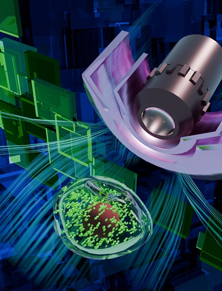
The study was published in the latest issue of ‘Opto-Electronic Advances’, a leading journal in the field of optics, and was selected as the cover feature.

▲ Cover of the latest issue of ‘Opto-Electronic Advances’: 3D schematic of a tissue stabilization device for subcellular organelle imaging


