


-
- Global AMC MENU
- NEWS
- HEALTH
- PEOPLE
- Introduction


Asan Medical Center Team Led by Professor Ji Hoon Kim Reaches 100 Robotic Liver Resections for Liver Cancer and Tumor Patients
Enhanced Precision through 3D Imaging and Fluorescence Guidance
Smaller Incisions, Less Pain, and Faster Recovery… With Advanced Imaging Support, as Safe as Open or Laparoscopic Surgery
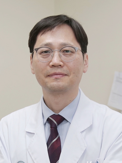
▲ Professor Ji Hoon Kim of the Division of Liver Transplantation and Hepatobiliary Surgery at Asan Medical Center
The liver is rich in blood vessels and has a complex anatomical structure, making resections highly prone to bleeding. For this reason, liver cancer surgeries have traditionally been performed through open or laparoscopic procedures to allow for immediate response in the event of massive bleeding.
Asan Medical Center is safely performing complex liver cancer resections with robotic surgery by using 3D imaging to visualize the patient’s liver structure and fluorescence guidance to clearly identify the area to be removed.
A team led by Professor Ji Hoon Kim of the Division of Liver Transplantation and Hepatobiliary Surgery at Asan Medical Center performs the highest number of robotic liver resections in Korea each year for patients with liver cancer or liver tumors, recently reaching 100 cases in just one year and five months.
Robotic liver resection is performed by inserting robotic arms through four small abdominal incisions, each about 8 millimeters in diameter. For surgeons, the system corrects hand tremors and provides a magnified surgical view up to ten times, reducing the risk of injury to major blood vessels. For patients, the procedure minimizes incisions, pain, and bleeding, lowers the risk of complications, and offers a faster recovery with shorter hospital stays.
Thanks to the many advantages of robotic surgery, it has already become a standard approach for various cancers such as prostate, rectal, and kidney cancers. The liver, however, has long been considered an organ where the wider use of robotic surgery is difficult to achieve.
The liver has a complex network of vessels such as the portal vein and hepatic veins, along with high blood flow and intricate biliary anatomy. In emergencies, rapid hemostasis is essential, but during robotic surgery it can be difficult to quickly convert to open surgery or achieve immediate bleeding control. In addition, because each patient’s liver anatomy is different, customized resections are often required. While robotic surgery offers excellent visualization, the inability to palpate directly makes it challenging to fully assess anatomical structures.
To overcome these limitations, Professor Ji Hoon Kim’s team has actively incorporated 3D imaging and fluorescence guidance into robotic liver resections. For every patient, two dimensional liver images are reconstructed into 3D views, enabling the surgical team to clearly identify each individual’s unique bile ducts, blood vessels, and portal structures and to perform precise resections at the segmental level.
At the same time, the team became the first in the world to introduce fluorescence imaging with indocyanine green (ICG) into robotic liver resection, now applying it to most patient procedures. After blocking the portal vein or segmental vessels, ICG is injected intravenously. The liver tissue with blood flow then glows green, while the blocked area shows no fluorescence. Because the resection margin can be seen clearly in real time, surgeons can accurately follow the disappearing fluorescent boundary, allowing for precise ‘detachment’ resections.
Conventional liver resections rely heavily on the surgeon’s anatomical knowledge, visual observation, and experience. When resection margins are unclear, there is a risk of incomplete tumor removal as well as unnecessary damage to healthy liver tissue. With ICG, however, surgeons can resect along natural boundaries as if gently detaching the tissue. This approach reduces the likelihood of residual tumors or excessive resection, helps preserve liver function, and minimizes injury to blood vessels and bile ducts, thereby lowering the risk of complications.
Patients who underwent robotic liver resection had an average hospital stay of 4 to 6 days, which was shorter than that of open surgery (about two weeks) or laparoscopic surgery (about one week).
Although liver cancer resection is considered one of the most complex procedures in surgery, Professor Ji Hoon Kim’s team has been safely performing robotic resections even for tumors larger than 10 centimeters by applying 3D imaging and ICG techniques.
Professor Ji Hoon Kim of the Division of Liver Transplantation and Hepatobiliary Surgery at Asan Medical Center said, “With the active use of advanced imaging support, robotic liver resection can be performed as safely as open or laparoscopic surgery. At Asan Medical Center, more than 100 robotic liver resections are expected to be performed this year. We hope that as the scope of robotic liver surgery expands, many liver cancer patients will be able to maintain a high quality of life after surgery.”
He added, “When tumors involve major liver vessels such as the hepatic veins or the portal hilum, open surgery may be the safer option. Therefore, we recommend that patients consult closely with specialists to determine the most appropriate surgical approach.”
Meanwhile, Professor Ji Hoon Kim’s team is also contributing to the training of the next generation of surgeons by providing education programs and regular workshops for liver transplantation specialists in Korea and abroad, offering hands-on observation of laparoscopic and robotic liver resections. This year alone, the team has conducted two live surgery demonstrations to share their expertise widely, further establishing Asan Medical Center as a leading center for robotic liver surgery.
ASAN MEDICAL CENTER
BROCHURE DOWNLOAD
Sharing your hope
ASAN MEDICAL CENTER(2019)
