


-
- Global AMC MENU
- NEWS
- HEALTH
- PEOPLE
- Introduction


AMC Medical Team’s Imaging Technology Recognized as the Most Significant Advancement in Nuclear Medicine at the SNMMI Annual Meeting
PET/CT Study on Deep Vein Thrombosis Recognized as the Only Selected Technology Among 1,500 Abstracts for Its Contribution to Advancing Nuclear Medicine
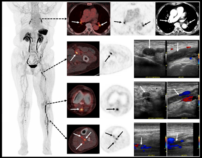
▲ A medical team from the Department of Nuclear Medicine and the Division of Vascular Surgery at Asan Medical Center received the “Image of the Year Award” from the Society of Nuclear Medicine and Molecular Imaging for their study on the “Evaluation of the Diagnostic Accuracy of Thrombus Specific Molecular Imaging Using 18F GP1 PET/CT in Patients with Acute Lower Extremity Deep Vein Thrombosis.” The image shows a 75 year old female patient’s 18F GP1 PET/CT scan, which simultaneously detected extensive deep vein thrombosis in the left femoral and calf veins and pulmonary embolism in both lungs.
A medical team from the Department of Nuclear Medicine and the Division of Vascular Surgery at Asan Medical Center recently received the “2026 SNMMI Henry N. Wagner, Jr., Image of the Year Award” at the Annual Meeting of the Society of Nuclear Medicine and Molecular Imaging held in Los Angeles, USA.
The “Image of the Year Award” is presented annually to the most innovative imaging technology among studies presented at the SNMMI Annual Meeting that has made significant contributions to the early detection and diagnosis of diseases, as well as treatment decision making.
A medical team from the Department of Nuclear Medicine and the Division of Vascular Surgery at Asan Medical Center received the “Image of the Year Award” in recognition of their research on the “Evaluation of the Diagnostic Accuracy of Thrombus Specific Molecular Imaging Using 18F GP1 PET/CT in Patients with Acute Lower Extremity Deep Vein Thrombosis.” The study was selected as the most outstanding research among approximately 1,500 abstracts submitted to the 2026 Annual Meeting of the Society of Nuclear Medicine and Molecular Imaging (SNMMI).
Founded in 1954, the Society of Nuclear Medicine and Molecular Imaging (SNMMI) is a leading international academic society dedicated to advancing nuclear medicine, molecular imaging, and theranostics. Experts in nuclear medicine and molecular imaging from around the world participate in the annual meeting to share the latest research achievements and discuss emerging technologies in depth.
To date, Asan Medical Center is the only institution in Korea whose researchers have received the “Image of the Year Award.” Thirty years after the Nuclear Medicine team at Asan Medical Center first received the award in 1996 for brain SPECT imaging using the dopamine transporter imaging radiopharmaceutical 99mTc TRODAT, the team has once again achieved this prestigious recognition.
Deep vein thrombosis (DVT) is a relatively common vascular disease in which blood clots form mainly in the veins of the legs, occurring in approximately 1 to 2 out of every 1,000 people. Early detection is crucial, as the clot can travel to the lungs and cause a potentially life threatening condition known as pulmonary embolism.
However, conventional imaging methods such as venous ultrasonography and CT require indirect findings, such as vascular compression or contrast filling defects, to estimate the presence of blood clots. In addition, these methods have limitations in evaluating certain areas, including the calf and pelvic veins.
A research team consisting of Professors Sangwon Han and Seung Jun Oh of the Department of Nuclear Medicine and Professor Jun Gyo Gwon of the Division of Vascular Surgery at Asan Medical Center successfully achieved direct visualization of blood clots using PET/CT with the radiotracer 18F GP1, which selectively binds to specific receptors on the surface of platelets involved in thrombus formation. The 18F GP1 PET/CT demonstrated high diagnostic accuracy and enabled simultaneous evaluation of both deep vein thrombosis and pulmonary embolism through a single imaging examination.
This study was highly recognized for demonstrating the potential of 18F GP1 PET/CT to detect thrombi in areas that are difficult to evaluate with conventional imaging methods, such as calf and pelvic veins. It also suggested the possibility of expanding the application of this technology beyond deep vein thrombosis to the diagnosis of various thrombotic diseases, including stroke and cardiovascular disease.
The study is particularly significant as a translational research project supported by the Research Oriented Hospital R&D Program of the Ministry of Health and Welfare. Through multidisciplinary collaboration combining the radiopharmaceutical development capabilities of the Department of Nuclear Medicine and the clinical expertise of the Division of Vascular Surgery at Asan Medical Center, the research successfully connected basic scientific achievements with actual patient care.
Professor Sangwon Han of the Department of Nuclear Medicine at Asan Medical Center said, “This study represents an achievement that has gained international recognition for the clinical value of thrombus specific PET imaging technology and demonstrates that new molecular imaging technologies can be successfully applied in real patient care. We will continue to conduct innovative research to accurately diagnose various thrombotic diseases and contribute to improving patient treatment outcomes.”
ASAN MEDICAL CENTER
BROCHURE DOWNLOAD
Sharing your hope
ASAN MEDICAL CENTER
