


-
- Global AMC MENU
- NEWS
- HEALTH
- PEOPLE
- Introduction


Professors Jun Ki Kim and Junyeop Lee of Asan Medical Center Develop Platform to Predict Treatment Response in Retinal Diseases
Highly Sensitive Biomarker Detection and AI Enable Intraocular Biomarker Analysis with 96% Diagnostic Accuracy
“Even with Small Sample Volumes, the Platform Could Identify Patients Less Likely to Respond to Treatment in Advance, Enabling Personalized Therapy.”
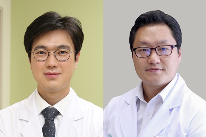
▲ (From left) A research team led by Professor Jun Ki Kim of the Department of Convergence Medicine and Professor Junyeop Lee of the Department of Ophthalmology at the University of Ulsan College of Medicine and Asan Medical Center
The three major eye diseases that can lead to blindness are age related macular degeneration, diabetic macular edema, and retinal vein occlusion. Although anti vascular endothelial growth factor (VEGF) injections, in which medication is directly injected into the eye, have recently been widely used for treatment, limitations remain, as approximately one in three patients shows a poor response or experiences delayed therapeutic effects.
Amid growing demand for technologies capable of predicting individual patients' responses to specific treatments in advance, a Korean research team has recently developed a technology that uses artificial intelligence to analyze biomarkers present in intraocular fluid, enabling accurate differentiation of major retinal diseases and prediction of treatment responses.
A research team led by Professor Jun Ki Kim of the Department of Convergence Medicine and Professor Junyeop Lee of the Department of Ophthalmology at the University of Ulsan College of Medicine and Asan Medical Center has developed an integrated diagnostic platform that combines highly sensitive biomarker detection technology with artificial intelligence to enable precise diagnosis of retinal diseases and early prediction of treatment responses. The platform achieved a diagnostic accuracy of 96.45%.
The technology is expected to contribute to the precise diagnosis of vision threatening eye diseases and the establishment of personalized treatment strategies for individual patients. The study was recently published in Materials Today Advances (five year impact factor: 10.0), a leading journal in materials science and convergence engineering.
Imaging modalities such as optical coherence tomography (OCT) and fundus photography are primarily used to diagnose retinal diseases and evaluate treatment outcomes. While these techniques can identify structural changes, such as retinal swelling or alterations in blood vessel morphology, they have limitations in the real time assessment of the underlying biological changes that drive disease progression. In particular, it often takes more than a month to determine whether a patient is responding to treatment, meaning that even non-responders may need to undergo repeated injections before treatment efficacy can be confirmed.
In contrast, biochemical analysis of disease related biomarkers enables direct assessment of biological changes. However, it requires at least 100 microliters (µL) of aqueous humor, making its clinical application challenging due to the relatively large sample volume required and the complexity of the analytical process. Aqueous humor is the clear fluid that fills the space between the cornea and the lens.
To address these limitations, the research team developed an integrated diagnostic platform that combines Surface Enhanced Raman Spectroscopy (SERS), a highly sensitive biomarker detection technology, with an artificial intelligence (AI) algorithm to enable early prediction of retinal diseases and treatment responses.
First, the research team developed its own gold coated zinc oxide (Au-ZnO) nanorod based chip. When an aqueous humor sample is applied to this nanostructure, biomolecules within the fluid infiltrate the gaps between the rod shaped nanorods. The structure acts as a "nano filter," effectively concentrating disease related biomolecules.
Using the SERS based diagnostic platform, the researchers further enhanced the concentration and detection of these biomolecules. By increasing sensitivity through the localized surface plasmon resonance (LSPR) mode of metallic materials, they amplified Raman signals by more than 300,000 fold. This enabled the team to obtain biochemical data in real time from less than 5 microliters (µL) of aqueous humor without the need for complex sample preprocessing.
Furthermore, the research team established a precision diagnostic system by applying artificial intelligence to the biochemical data obtained from aqueous humor samples. When tested in a cohort of 38 participants, including 12 normal controls (cataract patients without retinal disease) and 26 patients with retinal diseases, the system achieved a high accuracy of 96.45% in distinguishing the presence of retinal disease.
The AI based analysis algorithms, including the principal component linear discriminant analysis (PC-LDA) and principal component quadratic discriminant analysis (PC-QDA) models, also demonstrated accuracies of 87.63% and 86.45%, respectively, in differentiating specific types of retinal diseases. These models were able to precisely distinguish the biochemical patterns of complex retinal disorders, including age related macular degeneration, diabetic macular edema, and retinal vein occlusion.
In particular, the researchers highlighted the platform's ability to predict individual patients' responses to anti VEGF therapy as its most notable achievement. The integrated diagnostic platform developed by the team was able to detect biochemical changes within the eye that were not yet visible on post treatment imaging studies. It also identified treatment responders and non-responders in advance with an accuracy exceeding 90%. These findings provide a basis for identifying patients who are likely to benefit from treatment beforehand and for establishing personalized treatment strategies for patients at risk of blindness, enabling clinicians to determine the most appropriate therapy for each individual patient.
Professor Junyeop Lee of the Department of Ophthalmology at the University of Ulsan College of Medicine and Asan Medical Center said, “This study expands the conventional paradigm of ophthalmic diagnosis and treatment response prediction, which has primarily focused on identifying structural changes in the eye, to molecular level biochemical analysis. We expect that it will help identify patients with poor treatment responses at an early stage, establish personalized treatment strategies, and ultimately reduce the risk of blindness.”
Professor Jun Ki Kim of the Department of Convergence Medicine at the University of Ulsan College of Medicine and Asan Medical Center said, “By integrating nanophotonic technology with AI based algorithms, we were able to obtain meaningful biological information from extremely small samples of aqueous humor, which have traditionally been difficult to analyze. We will continue our research so that this noninvasive and cost effective diagnostic platform can be translated into clinical practice, including applications in screening for eye diseases and providing real time treatment guidance.”
Meanwhile, this study was supported by the Mid-Career Researcher Program of the National Research Foundation of Korea (NRF), funded by the Ministry of Science and ICT, as well as the Health Technology R&D Project and the Global Physician Scientist Training Program of the Korea Health Industry Development Institute (KHIDI), funded by the Ministry of Health and Welfare.
ASAN MEDICAL CENTER
BROCHURE DOWNLOAD
Sharing your hope
ASAN MEDICAL CENTER
